The Regional Brain
In this article I’m going to introduce the regions of the brain, the scientific terms for location, and the lobes that produce EEG signals that can be measured with a non-invasive device such as the OpenBCI.
This is the third post of my Background on the Brain series, which aims to help give amateurs the necessary background information to experiment effectively with the OpenBCI or a similar device. The previous two articles give my reasons for writing the series, and introduce the OpenBCI and some fundamentals of the brain. You can find them here:
Mastering Anatomical Terms for Location
It’s useful to understand the terms used to describe direction and locations in the skull, specifically the braincase or neurocranium.
To start, let’s tackle the scientific terms for a few regions of the brain that we’ll be learning more about. The terms used for relative position include:
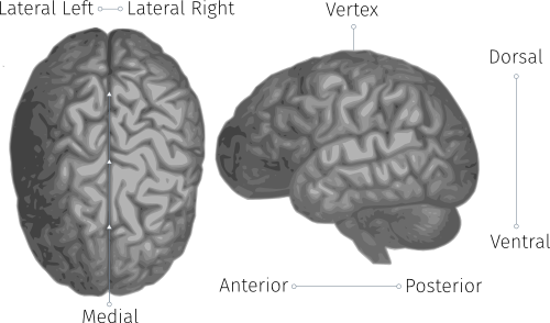
- Forward and Back:
- Anterior: toward the front of the head
- Posterior: toward the back of the head
- Vertex: central position
- Up and Down:
- Dorsal: toward the top of the head
- Ventral: toward the bottom of the head
- Superior: closer to the top (dorsal)
- Inferior: closer to the bottom (ventral)
- Right and Left:
- Medial: the mid-line of the head, running front to back
- Lateral Left: on the left side of the head
- Lateral Right: on the right side of the head
The Lobes of the Brain
The Cerebrum — mentioned in the last article as part of the forebrain, and described as the cap of a mushroom in the brain’s overall structure — makes up more than half of the brain’s total weight. The cerebrum controls all voluntary actions in the body with the assistance of the cerebellum, the tightly folded area at the back of the skull that’s responsible for coördinating movement. The cerebrum, mainly responsible for ‘high-level’ processing and decision making, has many connections to instinctive areas of the brain.
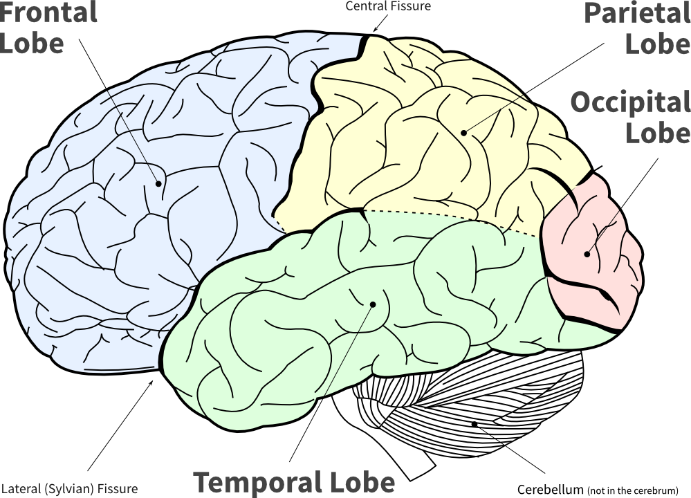
The cerebrum can be divided by basic anatomy into four regions: the frontal, occipital, temporal, and parietal lobes. The names of many brain regions are anatomical — for example, cerebellum is ‘little brain’ in Latin, and it resembles a small brain. At the time different parts of the brain were being named, their functions were not well understood. Naming them after anatomical characteristics (or who discovered them) was simple. Each of the four lobes is named after the bone that it’s under, so the imaginatively named frontal lobe sits under the frontal bone.
The lobes are divided by fissures. The central fissure separates the parietal lobe from the frontal lobe, the parieto-occipital fissure separates the parietal and occipital lobes, and the lateral fissure (also called the sylvian fissure) separates the parietal and temporal lobes. Additionally, the entire brain is divided into two hemispheres by the medial longitudinal fissure mentioned in the last article.
-
The Frontal Lobe is the most popularized lobe of the brain: the frontal lobe is often called the brain’s CEO, and everyone likes to read about celebrities. It’s belived to play a large part in executive planning, attention, character, social awareness, and motivation — making decisions, then delegating what areas of the brain carry out the selected task.
-
The Occipital Lobe is also well represented in popular literature, as the optic nerve runs from the eyes to the occipital lobe, where most of the visual processing is performed. The occipital lobe plays a key role in reading, writing, and spelling, as well as just about anything else involving object recognition, colour, and identification. When the eyes are closed, alpha waves from the occipital lobe are the strongest signal that can be picked up by an EEG. Measuring alpha activity with electrodes over the occipital lobe is a common first EEG measurement: the signal is easy to pick up, and you can see whether the eyes are open or closed from the EEG recording.
-
The Parietal Lobe is where inputs other than sight and sound are routed to and processed. Called somatosensory inputs, these include touch and temperature. The parietal lobe sits like a saddle at the back of the brain, behind the frontal lobe, and above and slightly forward of the occipital lobe. The name comes from the parietal bone, the section of the skull that the parietal lobe is under.
-
The Temporal Lobe is below the lateral fissure. It processes auditory input, including speech and language. The temporal lobe contains the hippocampus, and plays a key role in the formation of long-term memory.
The Cerebral Cortex
Cortex means the outermost layer of an organ in anatomy, and comes from the Latin for bark, rind, shell, or husk. The human cerebral cortex is the outermost layer of the brain, typically 2–4 millimetres thick, and is part of the cerebrum. The outer layer of the cerebellum is called the cerebellar cortex. The motor cortex and the prefrontal cortex are smaller areas within the cerebral cortex.
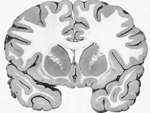
The cerebral cortex is of special interest because it is mainly grey matter. When you look at the cross-section of the brain, the outer layer appears grey, while inner regions are pinkish white, and called white matter. The colour difference is caused by the ratio between cell bodies (mainly neurons) and nerve fibres (axons) in the different areas: Grey matter has more cell bodies, while white matter has more nerve fibres (axons). White matter gets its colour from the white, fatty myelin sheath that forms around axons.
The cerebral cortex is folded into ridges, or Gyri, and furrows, Sulci, which significantly increases its surface area.

The cerebral cortex is present only in mammals, and because it’s the outermost layer of the brain, most EEG activity that can be picked up with an OpenBCI or similar device comes from the cerebral cortex.
Conclusion
Hopefully this article has introduced you to the terms for location in the brain, and given you a rough road map of the territory as you start experimenting with brain-computer interfaces and the OpenBCI. The next article will dive into brainwaves: what they are, where they come from, and their many varieties. You can subscribe to our RSS feed to get the rest of the articles in the series as they’re published.
If I’ve missed anything vital, or you have additional questions or comments, just leave a comment below and I’ll do my best to help out (or you can contact me directly).