OpenBCI Brain Basics: Structure and Biology
Understanding a little bit about what we know of the brain and how it works is a necessary step for anyone interested in brain-computer interfaces, EEG, or biofeedback. This article is an overview of some basics that make brain functions discussed later in the series easier to grasp.
This is the second article in my Background on the Brain series. If you haven’t read it, you can find the first post, OpenBCI: Background on the Brain, for an overview.
Contents:
- Introduction
- What we don’t know
- Neurons
- Cerebrospinal Fluid
- The Old and the New
- The Hemispheres
- Conclusion
What We Don’t Know
The brain remains to a large degree a mystery unto itself. Consider some of the things we don’t know about the brain: what consciousness is, for example. What’s the physical basis of subjective experience, cognition, and attention? These fundamental questions haven’t been answered! I recommend a look at Wikipedia’s list of Unsolved Problems in Neuroscience to keep what we don’t know in perspective. And I don’t know many things others do, so keep that in mind also.
Neurons
Neurons, or nerve cells, are one of the most important types of cells in the brain. Neurons process and transmit information through electrical and chemical signals, and there are currently estimated to be ~86 billion of them in an average human brain. For comparison, that’s 13 times more neurons in your brain than there are humans on earth.
A rather approximate and unattractive diagram of a Neuron.
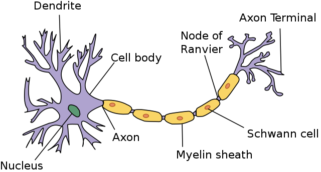
Neurons typically receive input though their dendrites (see diagram), process it in the cell body, and respond with electrochemical signals down their axon. Axons are also known of as nerve fibers, and the longest can reach a meter in length in humans — though most are far shorter.
 A more detailed diagram of a motor neuron
Synapses allow a neuron to pass electrical or chemical signals to another cell. Synapses between neurons usually connect the axon of one to a dendrite of another, but there are also synapses that connect dendrites to dendrites, and axons to axons.
Chemical synapses have a small (~20nm) gap between the axon terminals of one cell and the dendrites of another called the synaptic cleft. The sending (‘pre-synaptic’) cell releases neurotransmitter molecules into this gap, which bind to receptors on the other (‘post-synaptic’) cell. Chemical synapses can release multiple types of neurotransmitters, which can be inhibitory or excitatory.
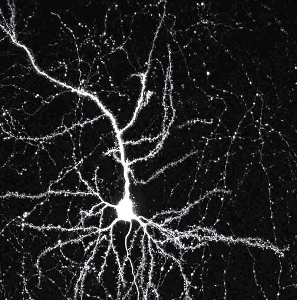 Image of a mouse neuron, made with a laser microscope. Photo Credit: Tony Pham, Baylor College of Medicine
Electrical synapses have a much smaller space of about 3.5nm between the two cells called a gap junction, where electrical signals pass between cells. Electrical synapses are faster, but they lack gain: the signal is always is the same or weaker in the receiving neuron. Neural systems that require the fastest possible response, such as defensive reflexes, often have electrical synapses. Another feature of electrical synapses: most of the time, the signals can be passed both ways, while chemical synapses are unidirectional.
Specialized types neurons perform specific functions. Sensory neurons, for example, respond to sensory stimuli — touch, sound, light, etc — and transmit signals to the brain. Motor neurons receive signals from the nervous system and initiate muscle contractions and other physiological processes.

Neurons are present in the body as well. Though the brain has higher concentration of them, there are large numbers of neurons in the spine, and in other organs such as the heart and intestines. In the spine, Spinal Interneurons take input from sensory neurons and relay it directly to motor neurons without sending the signal to the brain first; that’s how reflexes can be so fast, and how you can move your hand away from something that’s burning you without thinking about it: you don’t think about it until afterwards, if at all.
Neural Networks are formed by the connections between large numbers of neurons. Large numbers of neurons firing (transmitting signals down their axon) at once can generate ‘brainwaves’ that can be measured by an Electroencephalograph. The fifth article of the series, Brainwave Frequency Bands, will further explore brainwaves and their origins.
Cerebrospinal Fluid
As well as being protected by a thick skull, the brain is suspended in Cerebrospinal fluid, which provides further mechanical and immunological protection. Cerebrospinal fluid is often abbreviated CSF.

Cerebrospinal fluid is produced at rate of about half a liter a day, and flushed 4 times per day to clean out metabolites and toxins. It’s produced in the brain by the ventricular system, a set of four interconnected cavities (ventricles). Within each ventricle is a network of cells that produce the bulk of the CSF: the Choroid Plexus. The ventricular system is connected with the central canal of the spinal cord by the third ventricle, allowing for the flow of CSF to circulate.
The lymph system is responsible for clearing waste generated in most of the body, among other things, but it doesn’t extend to the brain. In the brain, instead of the lymph fluid, it’s cerebrospinal fluid that carries metabolic toxins and waste from the brain into the bloodstream.
The brain’s dense, and it’s hard for the fluid to circulate and get the waste out. According to a TED talk by neuroscientist Jeffrey Iliff, sleep plays an important (and surprising) role in the process. During sleep, more space is made for cerebrospinal fluid to circulate, and CSF flow increases. Furthermore, Iliff said “we discovered … the brain cells themselves seem to shrink, opening up spaces in between them,” making room for the cerebrospinal fluid to be pumped through the brain — on the outside of the blood vessels — and transport the day’s ‘waste’ from inside the brain’s tissue to the bloodstream!
Another benefit of CSF: neutral buoyancy. While the brain’s mass is about 3.3 pounds, or 1500 grams, when suspended in cerebrospinal fluid the brain’s net weight is equivalent to a mass of 25 grams. Existing in neutral buoyancy means the heavy, soft brain doesn’t cut off its blood supply by its own weight, as it would if it was resting on the skull rather than being suspended in cerebrospinal fluid.
The Old and the New
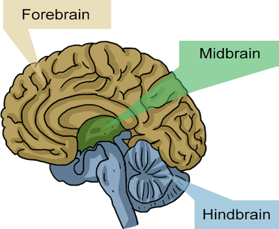 Image courtesy of Shmoop University
The brain can be considered as three sections divided by the type and complexity of their functions, with ‘higher-level’ processing farther from the brain stem. You can imagine the brain like a mushroom, where the hindbrain is the mushroom stem, with root into the spinal cord, the midbrain is the top of the stalk, and the forebrain surrounds the rest like a mushroom cap. It’s a primitive analogy, but gets the general idea of the three sections across.
-
The Hindbrain includes the upper part of the spinal cord, the brain stem, and the cerebellum. The hindbrain is believed to regulate vital functions such respiration and heart-rate. The cerebellum — latin for “little brain” — is a prominent region in the rear of the brain that contributes to coordination, precision, and accurate timing of motor control. The outer layer of the brain is folded in some areas, which greatly increases its surface area in the confined volume of the skull; the cerebellum is folded more tightly than other areas, which makes for a distinctive wrinkled appearance.
-
The Midbrain is located below the cerebral cortex, and above the hindbrain placing it near the center of the brain, and is associated with vision, hearing, motor control, sleep/wakefulness, and temperature regulation.
-
The Forebrain: is believed to have developed last in the brain’s evolution, and is the largest of the three areas. The forebrain controls voluntary movement, and enables higher intellectual functions such as speech and abstract thought, while older parts of the brain are responsible for subconscious tasks like the auto-regulation of bodily functions. The forebrain contains the Limbic system, which regulates the endocrines and is believed to play a key role in emotions, and the Cerebrum. Activity in the top layer of the Cerebrum, the Cerebral Cortex, is what’s normally picked up by an Electroencephalograph. The cerebral cortex also houses the much beloved Frontal lobes, often analogized as the brain’s chief executives.
Other ways of dividing the brain by regions and functions will be covered later in the series.
The Hemispheres
The medial longitudinal fissure — a deep groove running down the midline, front to back, of the brain — divides it into right and left hemispheres. The two hemispheres are connected by the corpus callosum, a wide band of neural fibers estimated to contain 200–250 million axons.
The two hemispheres are roughly mirror images of each other at the macroscopic level, with small differences, such a slight warping of the right side, bringing it just forward of the left side, called Yakovlevian torque. But the cellular composition of the cerebral cortex reveals that the types of cells, neurotransmitters, and receptors are can differ noticably between the hemispheres. Some of these differences are consistent across human beings — across species, even — but there are also observable differences from individual to individual.
My favorite source of highly scientific citations, Wikipedia, has an interesting observation about the generalizations made about the ‘right brain’ and the ‘left brain’:
Broad generalizations are often made in “pop” psychology about one side or the other having characteristic labels, such as "logical" for the left side or "creative" for the right. These labels are not supported by studies on lateralization, as lateralization does not add specialized usage from either hemisphere. Both hemispheres contribute to both kinds of processes, and experimental evidence provides little support for correlating the structural differences between the sides with such broadly-defined functional differences.
The extent of any modularity, or specialization of brain function by area, remains under investigation. If a specific region of the brain, or even an entire hemisphere, is injured or destroyed, its functions can sometimes be assumed by a neighboring region in the same hemisphere or the corresponding region in the other hemisphere, depending upon the area damaged and the patient's age. When injury interferes with pathways from one area to another, alternative (indirect) connections may develop to communicate information with detached areas, despite the inefficiencies.
But there is one thing about the hemispheres that “Pop” psychology has right: The left hemisphere of the brain controls the right half of the body, and vice versa.
Conclusion
I hope I’ve provided a useful overview of a few fundamentals of the brain, and that they’ll help provide a solid foundation for learning about the brain’s function on a macroscopic level. The next post will cover the terminology for location in the brain, and go over the regions of the cerrbral cortex and their functions, since the cerebral cortex generates much of the activity picked up by an Electroencephalograph such as the OpenBCI.
To hear when the next post is published (this one was somewhat delayed), you can subscribe to our RSS feed or follow me on Twitter. If I have omitted something, made an error, or you have a question feel free to reach out on twitter or by Email, or leave a comment below. Thanks for reading!